

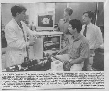
With these advances, the first demonstration of endoscopic OCT was reported in 1997 in the respiratory and gastrointestinal systems of a rabbit. The first demonstration of intravascular imaging was done in 1999, where blood had to be flushed out of the field (https://doi.org/10.1136/hrt.82.2.128). Dr. Brezinski directly compared OCT with IVUS, demonstrating the superiority of the former (https://doi.org/10.1136/hrt.77.5.397
). The first TD-OCT imaging catheter and system was commercialized by LightLab Imaging, Inc., formed in 1997 by Prof. Fujimoto, Mr. Swanson, and Dr. Brezinski. In 2001, Dr. Brezinski developed index matching to allow imaging through blood (https://doi.org/10.1161/01.CIR.103.15.1999). Intracoronary imaging in humans was achieve in 2003 by the Tearney and Bouma group (doi:10.1136/heart.89.3.317). Dr. Brezinski and Professor Fujimoto could not pursue in vivo human imaging because of COI. OCT systems began selling significantly in 2007. Lightlab Imaging is now owned by Abbott labs who has run double blind prospective trials showing morbidity and mortality benefits (DOI: 10.1056/NEJMoa2307770). "Among patients with complex coronary-artery bifurcation lesions, OCT-guided PCI was associated with a lower incidence of MACE at 2 years than angiography-guided PCI." A list of studies can be found on the Abbott web site. The only significant differences between the original LightLab system and those used in the clinical trials, besides using SS-OCT, is a rapid catheter attachment system to the OCT engine. Many advances which have been developed since 2000 which have not been incorporate in vivo by clinicians or in clinical trials. This includes PS-OCT, OCT elastography, and an OCT guidewire. Since 1993, he has advanced OCT through basic science, engineering, clinical trials, quantum physics, and classical physics. His major areas of OCT research are cardiology, osteoarthritis, quantum field theory of OCT, and adjuvant OCT techniques (PS-OCT, elastography, second order correlation spectroscopy).
Other Fields
Prior to 2000, Dr. Brezinski (with James Fujimoto PhD) would be the first to demonstrate OCT imaging for osteoarthritis (OA, discussed below), cervical cancer, esophageal cancer, bladder cancer, microsurgical guidance, breast cancer, prostate cancer, nerve repair, and tissue engineering.
OCT in the Diagnosis of Pre-Osteoarthritis (Pre-OA)
Dr. Brezinski has also done extensive work with OCT in diagnosing pre-OA, OA at reversible stages, in collaboration with orthopedic surgeon Scott Martin MD. This work spans more than 25 years. Initial work in the 1990s showed that OCT could diagnose early OA, but PS-OCT could diagnose pre-OA (PMID: 10090174)(PMID: 1140912). Pre-OA here is cartilage normal by visualization or MRI, believed to be at reversible stages. PS-OCT is identifying collagen disorganization. The first in vivo study was in patients undergoing knee replacement (https://doi.org/10.1186/ar1491). Dr. Brezinski would continue this work in the new millennium with most recently a double blind clinical trial in 2022 (https://doi.org/10.1016/j.ocarto.2022.100313). In this trial in patients undergoing arthroscopic meniscal surgery, abnormal PS-OCT predicted the onset of OA in 2.8 years.
OCT Work (2000-present)
General
After 2000, building his own OCT systems at Brigham and Women’s Hospital, he would go on to produce many of the most important OCT papers for the next 20 years, including inventing quantum second order correlation spectroscopy. Again, because of his IP (and owning LightLab), he could not participate in cardiac clinical trials because of COI. His collaboration with James Fujimoto PhD would be sporadic as they moved into different areas. He was continuously funded for more than 20 years, at times having five simultaneous NIH RO1s. He is the author of the Textbook of OCT where he wrote all chapters (physics, engineering, and clinical applications) except the clinical trials chapter (ISBN: 9780121335700).